What results would I expect to see in VEMP and vHIT in a patient with acute superior vestibular neuritis and acute inferior vestibular neuritis patient?
Vestibular neuritis is a condition where dizziness is caused due to an infection (mainly viral) of the vestibular nerve. The vestibular nerve has two branches which innervate the inner ear vestibular structures: The superior branch and the inferior branch. It is important not to forget that the neuritis can affect either branch individually or both branches at the same time. In order to understand the pattern of test results found in acute patients which have either superior vestibular neuritis or inferior vestibular neuritis it is important to know which organs each nerve synapses to.
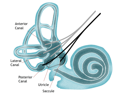
Have a look at the image below. The Superior vestibular (Grey) nerve connects to lateral semi-circular canal, anterior semi-circular canal and the utricle. Whereas the inferior vestibular (Black) nerve has connections to the saccule and the posterior semi-circular canal.
Now that we know the inner ear anatomy with relation to each vestibular nerve, we now need to know which piece anatomy each diagnostic test measures. The video head impulse test can measure the function of each of the semi-circular canal independently and their corresponding vestibular nerves, the cVEMP measures the function of the saccule and the inferior vestibular nerve and the oVEMP measures the function of mainly the utricle and the superior vestibular nerve. Therefore if a patient presents with a neuritis on the left superior nerve then you should expect the following results:
The Video head impulse test will show reduced VOR gain and catch up saccades in the lateral and anterior canals. Whereas the posterior vHIT should reveal normal test findings. The cVEMP will be normal as it only tests the inferior nerve but the oVEMP will be abnormal as the superior nerve needs to be intact to record a response from the utricle.
Related course: Balance testing for beginners
To assist you with differentiating between inferior vestibular neuritis and superior vestibular neuritis, see the tables below which show expected tests results in acute patients.
Direction fixed with fast phases toward the good ear
Reduces or is completely suppressed with fixation
Gaze-evoked nystagmus followng Alexander's law
Latency: within normal limits
Accuracy: within normal limits
Velocity: within normal limits
Evidence of spontaneous nystagmus may be present on tracings
Gain: within normal limits
Symmetry: within normal limits
Gain: enhanced when the stmulus moves in the same direction of the spontaneous fast phases
Total response: within normal limits
Unilateral weakness: less than 25%
Directional preponderance: less than 30%
Fixation index: less than 50%
Amplitude: reduced on affected ear or absent cVEMP
Amplitude: within normal limits
Catch-up saccades: not present or less than 50%
Gain: less than 0.7 on affected side
Asymmetry: larger than 7% toward affected side
Catch-up saccades: generated
Gain: typically reduced outside of the normative range
Phase: increased phase lead above normal range
Symmetry: asymmetry toward affected ear
Time constant: less than 10 seconds
Unilateral weakness: larger than 25% toward affected side
Directional preponderance: larger than 30% toward unaffected side
Amplitude: reduced on affected ear or absent oVEMP
This resource is a tool based on the needs of medical professionals and students that allows quick access to the typical assessment findings in a range of common vestibular disorders. The resource was developed to provide fast, easy-to-use, and always available information which can aid in reaching the correct diagnosis. The information contained within is provided as an information resource only, and should not be used as a substitute for professional diagnosis and management.
railCourse details
- Format: Reading
- Clinical area: Balance