Eclipse Support
The ocular vestibular evoked myogenic potential (oVEMP) is an evoked potential measured from the inferior oblique muscle and is used to assess the vestibular system. There is still some debate over the origin of the response (Piker et al., 2011), however, the oVEMP is dependent on the integrity of the superior vestibular nerve (Jacobson et al., 2011).
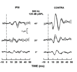
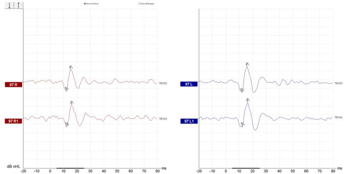
The oVEMP is recorded using surface electrodes at four sites on the face and an Amplitude Asymmetry Ratio is calculated to determine if the above-mentioned parts of the vestibular system are intact and working normally. The figure shows oVEMP recordings from a normal young adult (Murnane & Akin, 2009).
The oVEMP is a test used in addition to traditional vestibular testing (e.g., VNG) to assist in the assessment of vestibular function. oVEMP recordings provide valuable information to medical practitioners to assist them in the diagnosis of disorders such as Superior Semicircular Canal Dehiscence (SSCD) (Watters et al., 2006) and Meniere’s disease (Sandhu, 2012).
Patient preparation is very important. The electrode sites must be prepared and cleaned in order to obtain acceptably low skin impedances. It is recommended to have impedance values of 3kΩ or lower. The impedance value between each electrode should be balanced or similar in value.
The subject is either seated or in a reclined position and is instructed to maintain an upward gaze at 35 degrees for the duration of the recording (Kantner & Gürkov, 2014). Placing a static visual target on the wall or ceiling for the patient to look at during testing will ensure consistent activation of the inferior oblique muscle.
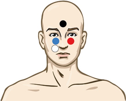
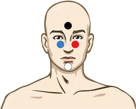
Use of this electrode montage does not require the active (white) electrode to be shifted during testing and is reported to provide large and replicable responses (Sandhu, 2013). Notice the reference electrode is placed slightly laterally from the orbital midline.
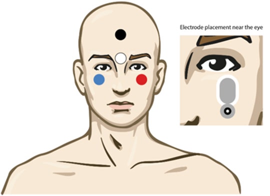
The reference electrodes should be placed as close as possible underneath the eye, slightly laterally in the orbital midline. Some clinicians prefer to clip the top part of the electrode to move it even closer to the inferior oblique. Avoid placement close to the medial canthus (inner corner of the eye) as a null-point exists where there is no oVEMP response present (Sandhu, George & Rea, 2013)
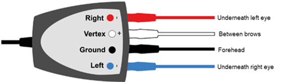
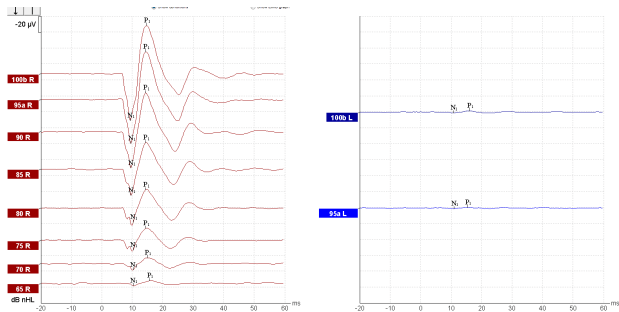
The oVEMP response is recorded from the inferior oblique muscle underneath the contralateral eye. Therefore, the right (red) electrode is placed under the left eye while the right ear is stimulated. Correct positioning of the electrode on the inferior oblique muscle is essential in obtaining a response (Sandhu, George & Rea, 2013).