Eclipse Support
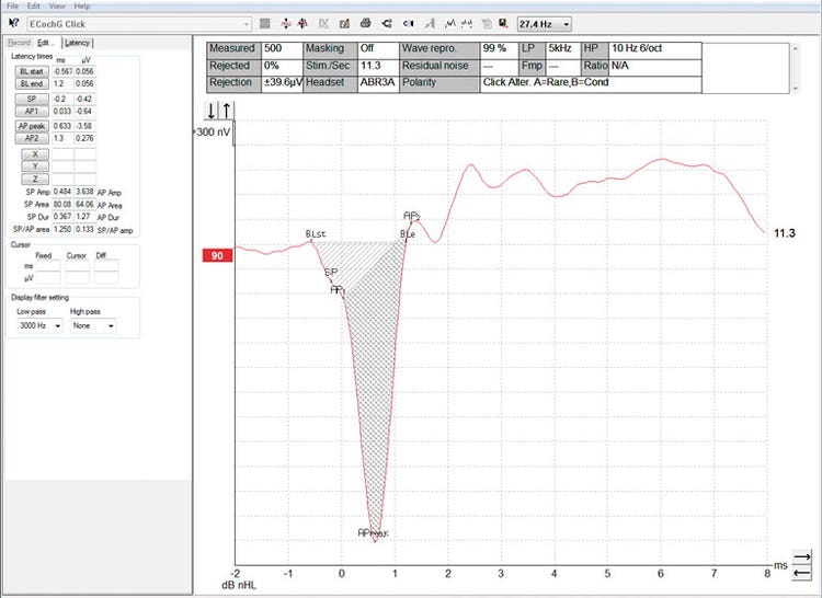
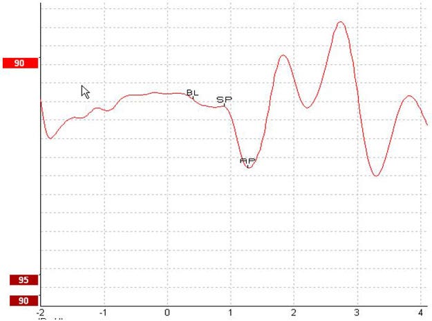
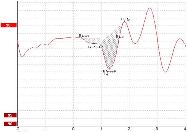
Electrocochleography, also abbreviated as ECochG or ECOG, is a measure of the electrical potentials of the cochlea. Typically, the measurement is characterized by the stimulus onset (baseline - BL), the response of the cochlea to the stimulus (summating potential - SP), and response to the synchronous firing of nerve fibers (action potential - AP). The AP is also known as Wave I.
The cochlear microphonic (CM) is also part of electrocochleography and has its own protocol. Measuring the CM requires slightly different test parameters than the SP and AP, why it is described in a separate guide.
See Figure 1 for an example of an electrocochleogram.
Electrocochleography can be helpful when diagnosing certain vestibular and auditory conditions. It is primarily used to diagnose Meniere’s disease and cochlear hydrops in particular. The SP and AP amplitudes, latencies, and their relationship are used to diagnose these conditions. Perilymphatic fistula (PLF), sudden hearing loss, and other pathologies may result in abnormal electrocochleography results. Superior canal dehiscence (SCD) may also result in elevated SP/AP ratios [1].
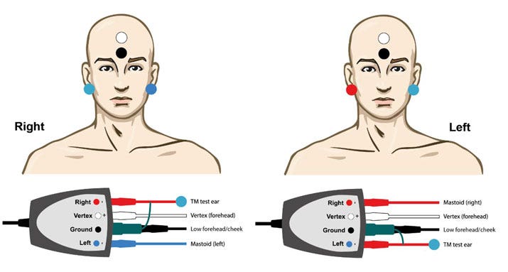
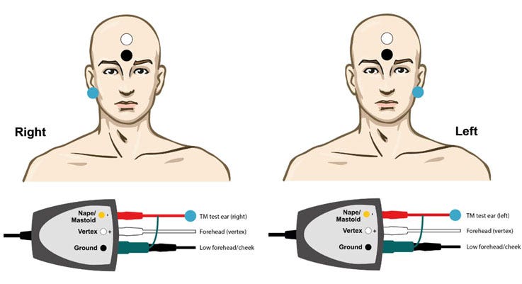
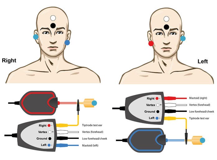
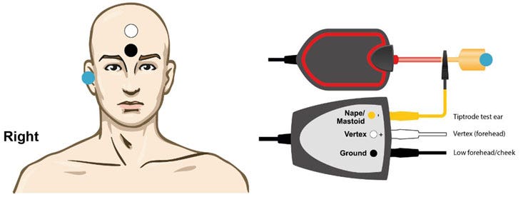
Surface electrodes are not adequate for electrocochleography. It is recommended to use TipTrodes, TM‑trodes or transtympanic electrodes to measure the electrocochleogram. Transtympanic electrodes will result in the most robust response but are not feasible for most clinics. Gold foil TipTrodes are sometimes used but TM-trodes will produce larger responses as they are closer to the site of generation.
Please observe the following before performing the test:
[1] Your patient should lie down and be relaxed or sleeping in a quiet environment.
[2] Examine the ear canal and tympanic membrane for any contraindications.